At the Rizzoli Institute a new personalised surgical treatment for ankle cartilage lesions
August 26th, 2021Osteochondral lesions of the ankle (i.e. lesions affecting the articular cartilage and the underlying bone, usually due to trauma), are often treated in a "biological" way in young patients: damaged tissues are regenerated by using the patient's own stem cells.
However, this option is only valid for young patients with a limited size of lesion. Biological cartilage reconstruction is no longer effective, with increasing age, in case of excessively large and deep lesions or failed surgical treatments.
On the other hand, total ankle joint replacement is used in elderly patients. Between these two groups of patients with such different age brackets there is a "grey area" of people for whom biological tissue regeneration is not indicated, but total ankle replacement is premature and excessive.
For these patients there are now new possibilities based on personalised surgery, which aims to cover and replace only the damaged area of articular cartilage and subchondral bone.

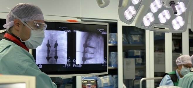
Prof. Stefano Zaffagnini, Director of the 2nd Orthopaedic and Traumatologic Clinic, and Massimiliano Mosca MD, Head of the Ankle and Foot Unit, in collaboration with Prof. Emeritus Niek van Dijk of the University of Amsterdam - AMC Hospital, have recently performed this innovative intervention at the Rizzoli Orthopaedic Institute on a patient suffering from a large and deep osteochondral lesion of the ankle who had already undergone a previous surgical treatment without success.

Using dedicated CT and MRI images of the ankle, a sort of "button" was made, a custom-made device with cobalt-chrome, titanium, hydroxyapatite alloy parts, which was implanted to replace the damaged cartilage and subchondral bone. The surgical instruments used for the operation were also designed and manufactured using 3D printing.